Lung Cancer Screening
Detection of lung diseases
Stellar Detector technology, combined with highly advanced iterative reconstruction algorithms such as SAFIRE4 and ADMIRE, enables the detection of lung diseases at an early stage using ultra low radiation doses.
- Collimation: 128 x 0.6 mTube settings: 100 kV, 10 eff. mAs
- Scan time: 1.9 sDLP: 15 mGy cm
- Scan length: 300 mmCTDIvol: 0.39 mGy
- Rotation time: 0.28 sEff. dose: 0.21 mSv
You can find out more information on the official Siemens website.
Advantages of computer-aided detection
When a chest CT arrives at his server, syngo.via immediately begins preprocessing, so the doctor has access to computer-assisted detection as soon as he opens the patient's file, and the results are immediately available in a PACS environment.
Early analysis of tumor endurance
syngo.CT Body Perfusion allows a detailed assessment of tumor endurance by analyzing its perfusion and vascularization to see at an early stage whether a patient is responding to therapy.
Take full advantage of virtualization
syngo.CT Liver Analysis allows you to try out different treatment options before the operation itself by even providing a virtual scalpel option.
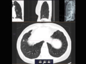
3D CT guided intervention with Adaptive 3D Interventional Suite
Interventions with complex anatomies (e.g., lung biopsies, liver ablations, and spinal surgeries) are best visualized with a 3D CT scan offered by the Adaptive 3D Interventional Suite with complete wireless control of movements and software options. Working in near real time, 3D offers a number of advantages: it allows examination of the entire organ using VRT and examination of the lesion with sagittal, coronal and axial MPRs. Smart automatic needle detection algorithms and trajectory planning tools allow doctors to always be on the right trajectory. Algorithms automatically select the optimal needle visibility, entry point and needle stitch angle. This 3D feature enables fast and accurate positioning in even the most complex anatomies, even from curved angles.
CT-guided 2D & 3D solutions that come with the SOMATOM Definition device family:
Adaptive 3D Intervention Suite
- The ultimate solution for 2D and 3D CT-guided interventions that require precision and speed
- i-Control offers complete control of software functionality and surface mobility (wired or wireless)
- Full 2D navigation capabilities
- 3D quantitative interventions
- Free work in all dimensions
- Reconstructed MPR views in near real time
- MPRs in coronal, sagittal, and oblique environments
- CT floroscopy, sequential and spiral scan modes
- Speed transition between i-Sequence, i-Spiral and i-Fluoro modes
- i-Fluoro CT floroscopy displayed in real time with up to 10 frames per second
- Intervention 3D toolbar enabled to support smart algorithms with syngo® 3D tools
- Automatic route planning by selecting the entry point and destination point
- Automatic needle detection
- Transition between patient view and needle view
- i-NeedleSharp technology to avoid needle-induced artifacts
syngo.CT DE Virtual Unenhanced
syngo.CT DE Virtual Unenhanced allows visualization of the concentration of contrast agent in the tissue. The basis of this approach is decomposition into iodine contrast agent, fat and liver tissue. Additionally, the app creates a virtual non-contrast display using Dual Energy information from fat and liver.
Clinical applications
Assessment of iodine accumulation in the abdominal lesion can be used as an indicator of whether the lesion is benign or malignant.
Features
- One-click virtual enhanced display calculation
- Slide bar that allows you to fine-tune the combined aspect ratio to see more enhanced or enhanced views
- Estimation of HU values and iodine concentration (mg / ml) in the virtual unenhanced or enhanced display
- Comparative view of virtual unenhanced display with combined data set from low and high kV data sets
- Collection of all findings and key views in the Findings Navigator during the analysis of an individual case
syngo.CT DE Lung Analysis
syngo.CT DE Lung Analysis application allows color coding of parts affected by for example pulmonary embolism and consequently shows a significantly lower concentration of iodine than unaffected vessels. It also allows rapid assessment of pulmonary perfusion defects and generates a lung image from an enhanced Dual Energy scan without the need for additional scanning.
The USPSTF recommends annual screening for low-dose CT lung cancer for patients between the ages of 55 and 80 who consume a minimum of 30 packs of cigarettes a year and continue to consume them as well as those who quit smoking less than 15 years ago. Learn more on the USPSTF official website.
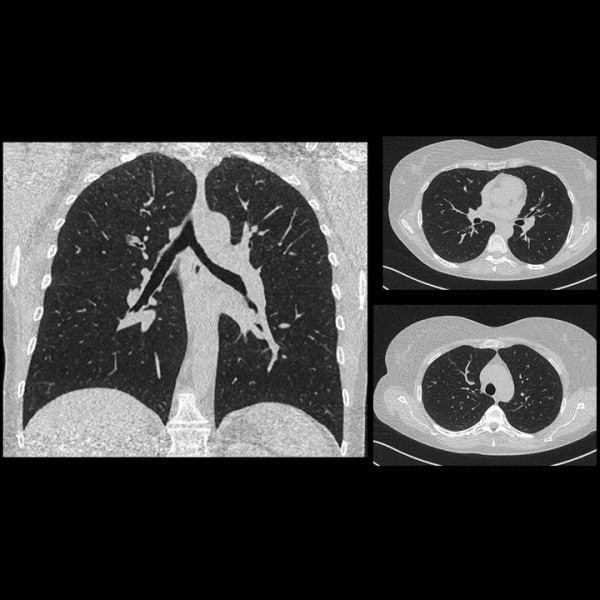
syngo LungCARE CT i syngo Lung CAD
syngo® LungCARE CT is designed to support physicians in confirming the presence or absence of pulmonary lesions, GGN of normal size, and GGO of smaller size. It allows volumetric analysis of pulmonary lesions and helps assess node changes during their growth.
3D CT guided intervention with Adaptive 3D Interventional Suite
Interventions of complex anatomies (e.g., lung biopsy, liver ablation, or spinal surgery) are best visualized with 3D CT guidance offered by the Adaptive 3D Interventional Suite. It allows free operation within a 3D rendered spiral or sequential CT data set. Full wireless bearing control and software functions are offered directly with the i-Control bearing.
Working in near real time, 3D offers many benefits - the entire organ can be seen using VRT and the lesion can be seen along with sagittal, coronal and axial MPRs. Smart algorithms for automatic needle detection and path planning tools ensure that the procedure goes in the right path. They automatically select the optimal viewing angle of the needle path, select the optimal entry point, and calculate the needle puncture angle. This 3D feature enables fast and accurate positioning in even the most complex anatomies, even from curved angles.
Main features
- i-Control: Complete control over stand movements and scanning software
- 3D CT guidance: Precise positioning in even the most complex anatomies with Adaptive 3D Interventional Suite
- 2D CT guidance: Ideal supplement for biopsy lavage and spinal injections
CT image navigation in minimally invasive therapy
Portable gantry CT scanners are playing an increasing role in hybrid ORs. Their versatility allows them to be used for a variety of minimally invasive interventions as well as for more invasive surgeries. The two-room configuration efficiently utilizes a single CT that can be moved between the CT intervention room and the angiosurgical space allowing for high utilization of the CT device. Space can be saved with a movable gantry that combines an angio and CT system in one room.
syngo Volume Perfusion CT Body
syngo Volume Perfusion CT Body enables quantitative 3D evaluation of dynamic CT data of organs and tumors by providing insight into blood flow and blood volume. syngo Volume Perfusion CT Body allows the assessment of perfusion disturbances and perfusion changes during therapy. It is especially useful in the differential diagnosis and monitoring of tumors.
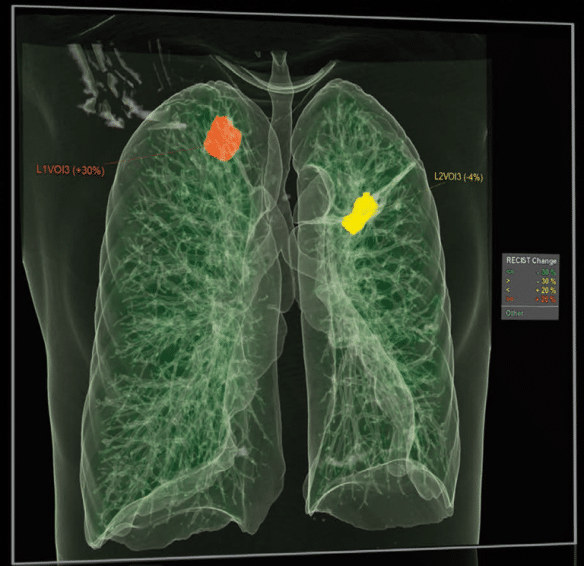
CT Oncology Engine
Cross-Timepoint Evaluation provides concise follow-up information about a patient's response to oncological treatment by calculating and displaying relevant visual changes over a period of time. Based on this, RECIST, size or any other parameter can be compared and changes in tumor size can be seen very easily.
CT Oncology applications provide professional analysis tools so that oncology professionals can get the most out of each CT data set.
Advantages of computer-aided detection
When a chest CT arrives at the server, syngo.via immediately starts preprocessing.
Since syngo.CT Lung computer-aided detection is available immediately at the syngo.via workplace, these results are available as soon as the patient file is opened. Thanks to Rapid Results technology, these results are immediately available in the PACS environment.
Early analysis of tumor endurance
Better treatment outcomes can be achieved with early therapeutic assessments. syngo.CT Body Perfusion allows a detailed evaluation of the endurance of the tumor by examining its perfusion and vascularization in order to be able to conclude at an early stage whether the patient responds to therapy.
Take full advantage of virtualization
The quality of medical care can be improved by virtual liver resection planning. syngo.CT Liver Analysis allows physicians to try several different approaches and accurately measure resection sizes before surgery itself using even the virtual scalpel available to them. The virtual plan can then be shared with other surgeons for analysis.