MRI Device Siemens MAGNETOM RT Pro edition Skyra 3T
- Field strength 3 Tesla
- Bore size 70 cm Open Bore design
- System length 173 cm
- System weight (in operation) 7.3 tons
- Minimum room size 31 m²
- RF Tim [204 x 48] [204 x 64] [204 x 128]
- Gradient strength XQ Gradients (45 mT/m @ 200 T/m/s)
- Helium consumption Zero Helium boil-off technology
In Radiochirurgia Zagreb clinic we are using MRI device Siemens Magnetom RT Pro edition Skyra 3T that is specifically adjusted for oncology to diagnose tumors in all stages.
syngo.MR OncoCare as part of the syngo.MR Oncology workflows Workflow enables accurate monitoring of oncological lesions, helping determine whether treatment is effective or potentially needs to be reconsidered. A comparison of up to eight exams at different time points is now possible, visualizing regional changes of tissue characteristics.
As an extension of the MR Oncology workflows, syngo.MR OncoCare, provides rich feature sets for histogram analysis and trending over multiple time points:
- VOI- and ROI-based histogram analysis
- Intuitive color definition for three histogram domains
- Savable presets for histogram analysis
- Back-mapping of histogram colors on the image of a reference
- Export in .CSV format of pixel intensity values
- Trending plot
- Improved communication towards referrers and oncologists with the back-mapping functionality
- Additional research possibilities with the export functionality
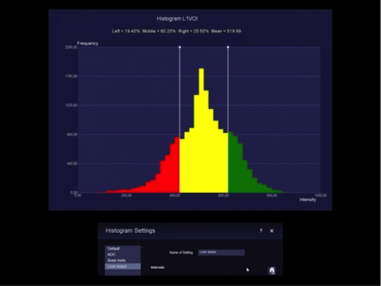
Histograms: Here, red indicates low ADC coefficient (area of concern), green high ADC coefficient (not of concern).
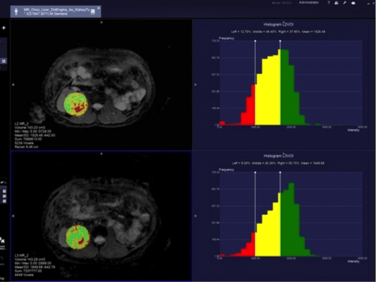
Back-mapping: The user-defined volume of interest can be “back-mapped” with colors that correlate to the histogram values. Here the patient’s histograms indicate a growth in area of concern from first exam (8% lower row) to follow-up exam (11% upper row), indicating that this patient may not be responding to therapy.
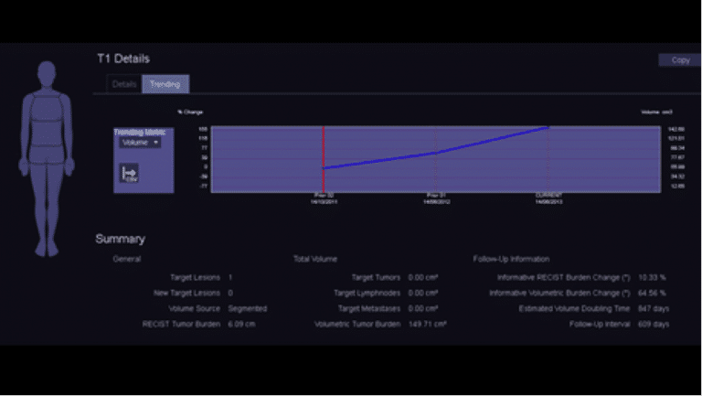
Trending plots: can be saved and included in structured reports.
Clinical Application
syngo.MR Onco Engine combines features that allow for efficient oncological reading and reporting.
Included Workflows: Onco Multi-Region, Onco Brain, Onco Liver, Onco TimCT
syngo.MR 3D Lesion Segmentation provides convenient volumetric evaluation of lesions
Onco Reporting: Each workflow contains a dedicated reporting template and oncological findings classification. Response calculations comply with RECIST and WHO Response Criteria
Additional Information
syngo.MR Onco workflows structure large amounts of data automatically and quickly into layouts focused on oncology reading. Each workflow contains a dedicated follow-up reading layout (optimized for dual monitor support). The 3D Lesion Segmentation is particularly useful for oncology applications (e.g. volumetric evaluation of tumors, lymph nodes and metastases), but also for non-oncology lesions with sufficient contrast to surrounding tissue. Intuitive editing tools allow adjustment to the segmentation if necessary. The perfect match to TimCT Onco and the syngo.MR Onco Engine enables an optimized workflow from scanning, to processing, to reading.
Oncology
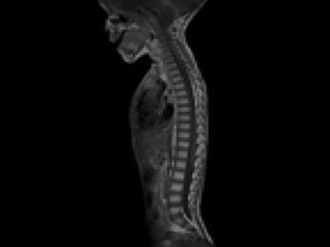
The Onco Suite MR device contains a collection of sequences, protocols and measurement tools that lead through a detailed assessment of a range of oncological conditions. It allows a full body scan for metastases in a single continuous motion with the TimCT Onco Dot Engine system.
Together with integrated management, this innovative technology improves patient throughput and provides great display quality.
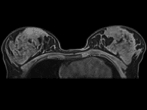
Health of women
The Team Coil interface is powered by a multitude of chest spirals that result in flexible breast display options to suit the diverse needs of patients. From a clinical presentation to help directing a biopsy, MAGNETOM Skyra offers a wide selection of chest coils and provides excellent display quality. Additionally, the Open Bore (70 cm) contained in the Skyra MAGNET enables comfortable reception of more patients. Breast Dot Engine offers different workflows for optimized display quality and consistency.
Find out more about breast magnetic resonance imaging here.
Health of men
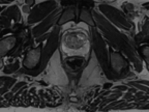
With the help of high-density body and spiral spirals or in combination with the endorectal spiral, the MAGNET Skyra provides an excellent multiparametric representation of the prostate in terms of morphology, physiology and function. Together with special solutions for analysis and reports, the workflow for prostate MRI is comprehensive.
Find out more about prostate magnetic resonance imaging here.
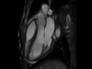
Cardiology
The Cardiology Package contains comprehensive 2D routine cardiology applications ranging from morphology and ventricular function to tissue characterization. Dot’s built-in navigation and increased Tim 4G SNR helps physicians make cardiology part of the clinical routine. The Cardiac Dot Engine helps eliminate one of the biggest challenges in cardiac MR by simplifying complete cardiac localization in just a few clicks.
Neurology
MAGNETOM Skyra offers high resolution and speed protocols even for patients who are not fully cooperative in examination. MAGNETOM Skyra Neuro package includes advanced protocols for diffusion and perfusion and fMRI. They are combined with Tim 4G coils with high element density and the latest iPAT2 reconstruction techniques that help provide fast display and excellent SNR.
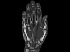
Orthopedy
The Ortho package is a comprehensive collection of protocols for examining joints including the spine. The new Tim 4G Ultra High Density Coils for MSK Review offer enhanced SNR and anatomical coverage. Dot’s customizable workflows include AutoAlign and AutoCoverage for the knee, hip and shoulder. Advanced syngo WARP techniques offer the acceptability of artifact reduction functionality (such as MR conditional implants) and MPR planning for high-speed 3D measurements Inline 3D multiplanar reformatting that helps workflow efficiency.
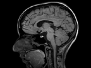
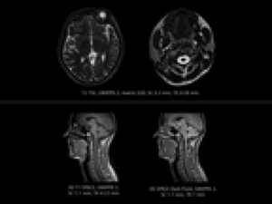
Brain, head and neck
Whether it’s brain changes or intracranial metastases, MRI helps make key clues for treatment planning with either IMRT or stereotactic radiosurgery. With the RT Dot engine, Siemens has produced special radiotherapy protocols for examinations of the brain and head and neck. These protocols are optimized for display during treatment with the help of flexible Tim 4G coils.
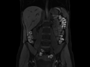
Body
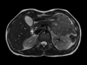
The 4G team enables high-channel body display thanks to the Body 18 and Spine 32 combination. MAGNETOM Skyra is also equipped with its own body imaging applications and provides ultra-fast high-resolution 2D and 3D protocols for the abdomen, pelvis, MR colonography, dynamic renal MRCP and MR utography. Abdomen Dot Engine also comes with standardized, efficient and comprehensive upper abdominal workflows with excellent display quality.
Prostate
In prostate radiotherapy, organ size is often misjudged using CT alone. MR with its soft tissue contrast allows for more precise delineation of the prostate and surrounding organs. With the use of multiparameter MRI (diffusion-weighted imaging and spectroscopy), you can even see metabolically active areas of the tumor, which helps identify areas to increase the dose and allows for better tailored treatment planning for better results.
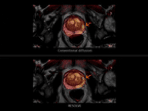
With RESOLVE, the diffusion-weighted display mode offers much better visibility to the magnet-prostate-2 changes and display quality, and common interference is reduced by a factor of three.
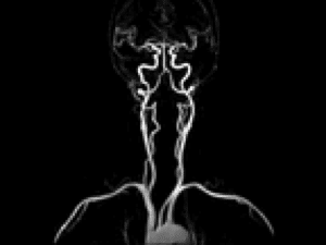
Angiography
Using the Angio Suite that comes with the Skyra MAGNET, excellent MR angiography can be performed to show arteries and veins with or without the use of a contrast agent. For contrast displays, the Angio Dot Engine helps to achieve optimized bolus timing at each review. Real-time views and the AutoVoiceCommands feature help the user set the optimal breathing time, scan and contrast medium.
Pediatrics
The time required for tissue relaxation in pediatrics differs greatly from the time required in the case of adults. Reasons for this are tissue developing, body size, faster heart rate, and level of cooperation during the examination. Protocols can also be customized to examine newborns.
StarVIBE
One of the most challenging groups for liver exams is the growing patient population unable to hold their breath. Dynamic imaging usually requires breath-holds, but many children, elderly or very sick patients cannot hold their breath for the required time or at all.
StarVIBE now delivers robust, free-breathing, and contrast-enhanced exams for these patients by intelligently resisting motion artifacts.
TWIST-VIBE
Always the right contrast in dynamic liver imaging.
Dynamic liver imaging remains a contrast-enhanced technique. Using contrast agent makes it necessary to catch the right point of the arterial phase. If this point in time is missed, crucial arterial information within the liver remains unseen.
TWIST-VIBE offers high temporal and spatial resolution with full 4D coverage for multi-arterial imaging with 100% precise contrast-timing.
Key benefits:
- Robust and fast liver imaging with full 4D coverage
- Excellent images to plan surgical intervention
- Reliable imaging from the very first shot
- Time and cost savings
- No need for rescans
LiverLab
LiverLab allows fat & iron evaluation and can be performed totally non-invasively. This allows trending and/or monitoring of patients suspicious of liver diseases especially in early disease stages.
The iron and fat values of the liver are important indicators for a variety of serious illnesses. Early evaluation could be a decisive step to better monitor early stages of diffuse liver diseases such as steatosis and hemochromatosis.
LiverLab is robust and fast enough to be implemented in routine clinical imaging with only a few clicks.
Elastography
Liver fibrosis with its associated cirrhosis and hypertension traditionally requires biopsy for diagnosis. MR Elastography (MRE) offers the possibility to evaluate relative liver stiffness noninvasively.
syngo GRACE
syngo GRACE (GeneRAlized breast speCtroscopy Exam) is a Single Voxel Spectroscopy technique optimized for breast spectroscopy.
Clinical Applications:
- Additional information in characterization of breast lesions seen in MR leading to better diagnosis
- May aid to improve the specificity for tumor diagnoses
- Follow-up of breast lesions after chemotherapy
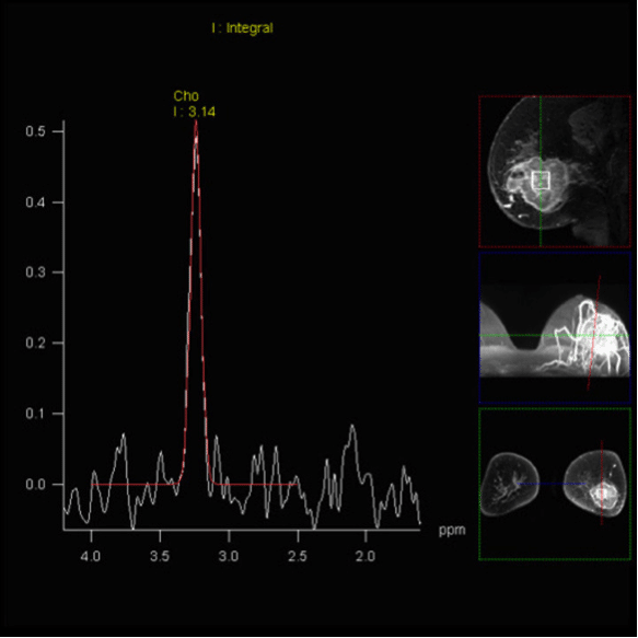
Increased Choline-signal within breast tumor, shown with syngo GRACE.
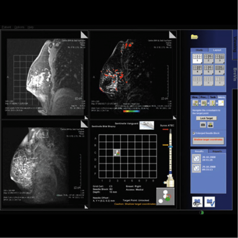
BreVis Biopsy
BreVis Biopsy is a professional solution for a fast and accurate MR biopsy workflow with automatic calculation of target coordinates. The user interface offers a guide for MR interventional planning and supports breast biopsies, i.e. Sentinelle Vanguard and related accessories. The easy-to-handle workflow enables shorter examination times for both patient and operator.
syngo RESOLVE
syngo RESOLVE (readout segmentation of long variable echo-trains) is a revolutionary new approach for obtaining high quality DWI images even in body regions which are strongly affected by susceptibility artifacts. It is not only largely free of distortions, but can also deliver sharp imaging at higher spatial resolution. syngo RESOLVE is especially attractive for the evaluation of smaller lesions in a wide range of DWI and DTI examinations.
Main application areas: brain, spine, breast and prostate imaging.
MRI enables excellent soft tissue contrast without using the dose, making it ideal for radiotherapy where the information gathered using CT could be enriched with valuable MRI information. Advanced imaging sequences such as RESOLVE and MR spectroscopy provide additional pathological characterizations, and using fast isotropic 3D sequences like SPACE and VIBE it is possible to achieve better anatomical correlation.
Here you can find more about the Siemens Magnetom RT Pro edition Skyra 3T MRI device.